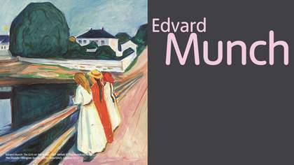
The exact nature of Munch’s haemorrhage is not known. However, we do know that he consulted Professor Johan Raeder (1889–1956), who was one of the most eminent ophthalmologists in Norway. Although no office records are available, Raeder referred to the artist’s illness in some later correspondence: ‘Munch… suffered from a period of over-exertion and of a generally reduced health… As a consequence… there occurred a very serious haemorrhage in his right eye… The haemorrhage brought about a very strong reduction in his eyesight, and this was all the more serious as vision in his left eye has always been less good. As it turns out, it was the eye he had always used when working that was affected.’
Raeder prescribed isolation and rest, and gave his patient the following note to ward off potential visitors: ‘Oslo, 10 May 1930. Herr painter Edvard Munch suffers from an acute eye disease caused by long-standing over-exertion. He needs complete bodily and mental rest for a long period of time. Any disturbance, oral, written, by telephone, or by telegraph, is to be entirely avoided.’
The ophthalmologist’s correspondence shows that the artist had a similar haemorrhage some years later in his left eye: ‘Mr Edvard Munch suffered six years ago from a very serious haemorrhage in his right eye. There now has occurred a similar condition in his left eye.’ This strongly suggests Munch may have had an underlying systemic disease that predisposed him to ocular haemorrhage. However, there is no documentary evidence to identify such an illness, and the second haemorrhage may not have bothered him visually since it was in his ‘bad’ eye. We know that he returned to painting, photography and correspondence after his 1930 haemorrhage, but Raeder may have been aware of some residual damage or been worried about recurrence.

Edvard Munch Disturbed Vision 1930
Oil on canvas
80 x 64 cm
© Munch Museum/ Munch-Ellingsend Group/DACS 2012
Courtesy Munch Musem Oslo
Munch drew several types of images during his convalescence. A recurrent one is a set of concentric circles, often vividly coloured, which resemble the aura that one sees around bright lights on a foggy day. It is possible that these represent a view through his resolving haemorrhage as he looked towards an electric light or the sun. He annotated many of his drawings ‘electric light’, ‘sunshine’, etc, to indicate the conditions under which they were made, but did not actually date them. The order of colours varies, so they don’t appear to be illustrating a rainbow effect, which would be constant. Whatever else, they do show that Munch must have been intrigued by the patterns of light and colour that suffused his eye as the haemorrhage slowly cleared.
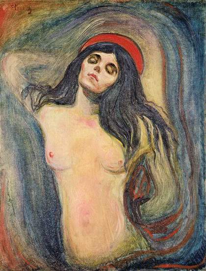
Other sketches documented the presence of a dense blind spot (scotoma) near the centre of his visual field. In one he even drew an arrow to show his point of fixation near the top of the dark shadow. In other diagrams the shadow was superimposed on sketches of people or a room, appearing in the lower or middle part of the scene. Sometimes it was simply an opaque shape, but in one striking watercolour it became a skull covering the foot of the artist’s bed. In that picture he portrayed himself with a hand covering his left eye, the better to observe the nature of the scotoma on the right.
This configuration is interesting, because Munch is known to have painted self-portraits using a mirror – in which case a hand over the left eye would have appeared to be over the right eye in the picture. One may presume that these sketches during convalescence were made to portray a subject, and were not mirror portraits. In a particularly poignant sketch, a nude figure is obscured partially by the irregular scotoma, expressing, it seems, Munch’s frustration as an artist who cannot see the core of his subject.

Edvard Munch The Artist’s Injured Eye (and a figure of a bird’s head) 1930–1
Crayon on paper
50.2 x 31.5 cm
© Munch Museum/ Munch-Ellingsend Group/ DACS 2012
Courtesy Munch Museum, Oslo
As the haemorrhage cleared further, he perceived misty, fibrillar shadows that sometimes took the form of a bird’s head and wings. Writing with respect to one of these drawings, he said: ‘The distance between the bird’s beak and the new beak beneath seems longer – I can make out two letters while earlier just one – the bird’s neck seems longer – I am clearing up on the left side.’ As the healing process continued, he described the debris within his eye: ‘There are dark spots which show up like small flocks of crows far up when I look at the sky – can they be blood clots which have been resting in the periphery of the damaged circular part – which by a sudden movement or by the effect of sharp light are moved from their origin – when they suddenly disappear it looks as if they fly down to their first place.’
Munch was deeply frightened by the ocular haemorrhage. He feared for his life, as well as his sight. This is evident in the portrait of the artist in bed with the death’s head scotoma, and in a drawing where he himself is portrayed with a skull-like visage. In another drawing his hands are held to his head in the same pose of fear and anguish as the famous subject of The Scream. The ominous bird appeared in a number of sketches, sometimes migrating from its inferior scotomatous position; in one image it was superimposed against a huge luminous sun. Munch was so worried about whether a return to painting would be possible that he spent a great deal of time during his convalescence working with photography, which he thought made fewer demands on his vision. However, by 1931 he was again painting regularly, and the strange images of birds and blind spots disappeared forever from his work.

Edvard Munch The Artist with a skull: Optical Illusion from the Eye Disease 1930
© Munch Museum/Munch-Ellingsend Group/DACS 2012
Courtesy Munch Museum Oslo
A careful, almost compulsive observer, Munch documented the conditions for his photographic work and was very interested in the optics of that discipline. He also drew sketches of the shape of his blind spot, including one that shows exactly how words and letters were broken up and distorted by the scotoma. Accompanying this was the notation: ‘In the shadow of full sunshine after the eye has been covered, reading distance, spectacle lens #5.’ Others carried details such as: ‘Bedroom, electric light, no glasses, ½ metre.’ He measured the scotoma relative to a distant pole: ‘40 metres, eye fixed on top of pole but it is invisible.’ This precise and almost clinical interest in his disease led him to develop a grid technique for measuring and monitoring his scotomas.
Munch recovered from his ocular haemorrhage and produced some poignant portraits of himself as an old man, which show the same style and surprising colours of his earlier work. His eye disease in 1930 represented only a brief interruption of his long career, unlike Mary Cassatt, Edgar Degas, or Georgia O’Keeffe, all of whom eventually stopped painting because of poor vision. However, although the effects of ophthalmic problems such as macular disease and cataracts can be seen in the works of artists such as Degas and Monet, Munch was unique because he gave us scenes from within the eye itself. As ophthalmologists, we can recognise in his sketches not only the characteristics of floaters and ocular haemorrhage, but his efforts to document and measure the impact of his disease.